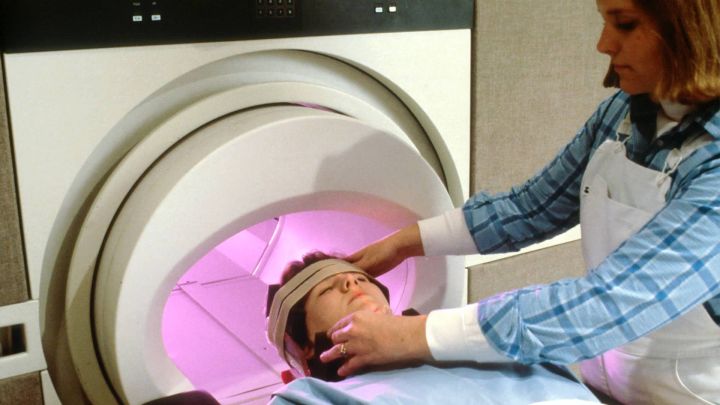
PET stands for Positron Emission Tomography and is known as "PET", an advanced nuclear medicine imaging technique; CT is short for Computed Tomography, an X-ray tomography technique that has been widely used in clinical practice and is still rapidly evolving. PET/CT ("PET CT") is the result of organically integrating these two technologies into the same machine and displaying images of different nature in the same machine.

What is the principle of PET/CT?
It is well known that malignant tumour cells are "robbers" in the body. They are very metabolically active and take in nutrients predatorily, and glucose is one of the main sources of energy for the body's cells (including tumour cells). As a result, malignant tumours take up far more glucose than other normal tissues. Based on this property, the injection of radionuclide-labelled glucose as an imaging agent (i.e. 18F-FDG) into the body can cause it to concentrate in diseased tissues such as tumours, thus presenting a bright spot in the image, and this concentration is quantified in PET/CT by the SUV maximum, whose full name is Standard Uptake Value (SUV ), is a semi-quantitative indicator commonly used in PET/CT for tumour diagnosis and is of greater value when evaluating the efficacy of treatment.
PET/CT imaging is like putting a GPS tracker on a bad guy, so that wherever he goes, he can be successfully located in a sea of people.

What is the difference between PET/CT and PET/MRI?
PET/CT and PET/MRI each have their own advantages and one test cannot replace all of them. According to Shi Huazheng, the latest force dual-source CT, for example, has unparalleled advantages in coronary artery imaging, as it can be used for non-invasive diagnosis, requiring only ¼ second to scan a lesion accurately, and the patient "eats" an extremely low dose of radiation, equivalent to that of two chest films (less than 1 mSv). The reconstructed image can be as thin as 0.16mm/layer, which is the level of vivisection and allows a very clear visualisation of the stenosis and the extent of the lesion, which is of great help to clinical diagnosis.
In addition, PET/CT and PET/MRI, the world's most advanced imaging screening devices, have significant advantages in the early screening and diagnosis of a wide range of benign and malignant tumours throughout the body, the nervous system and the cardiovascular system, and can detect tumours as small as millimetres, which is particularly useful for the early detection and treatment of liver cancer, breast cancer and pancreatic cancer.

